Software Vendors & OEMs
Ready to integrate web DICOM PACS Viewer and connectivity solutions

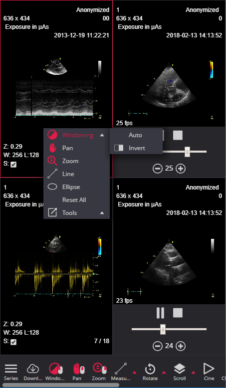
DICOM
ECG VIEWER
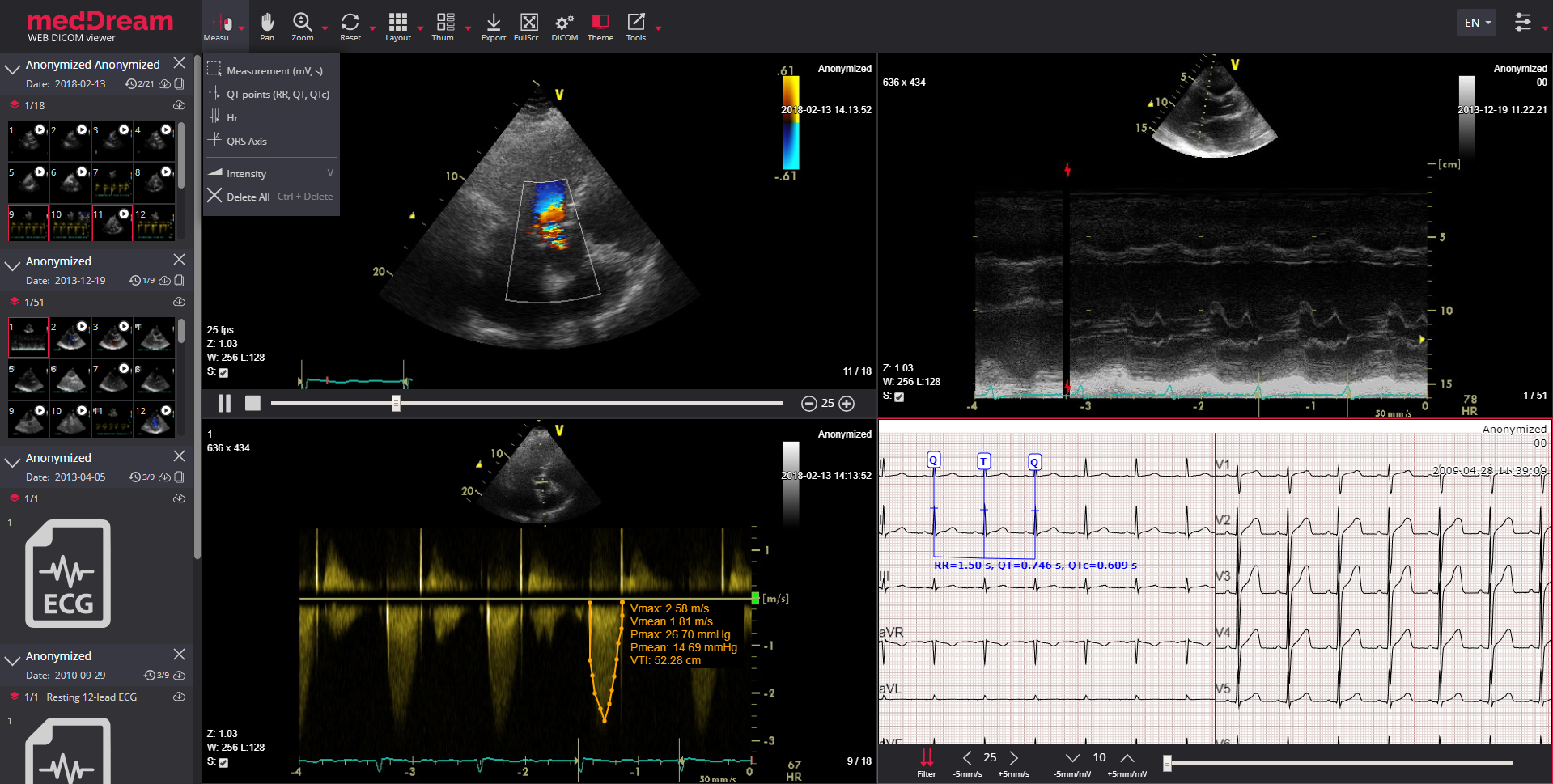
MedDream DICOM ECG Viewer provides standard cardiology image manipulation tools, but also a way to read, manipulate and interpret electrocardiography data. DICOM ECG & Ultrasound Viewer allows to zoom, measure and quantify the ECG dicom and US dicom data.

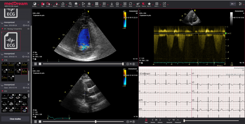


The MedDream DICOM Viewer provides not only standard image manipulation tools, but also a way to read, manipulate and interpret electrocardiography (ECG) and ultrasound (US) DICOM data.
DICOM ECG Viewer’s manipulation tools are all presented in an innovative zoom model, which allows zooming in/out, measure and quantify the ECG data:

Measurement (mV, s). Area calculation indicating beats per minute, time, millivolt (mV, s, bpm);

QT points (RR, QT, QTc). QT interval - the RR interval is calculated as well as QT and the QTc (based on Bazett’s formula);

HR. Measure heart rate (HR) and compare its interval variance over the ECG;

QRS axis. Measure the QRS electrical heart axis;

Studies comparison. Compare of two or more ECGs by normalizing and then overlaying them on one another.
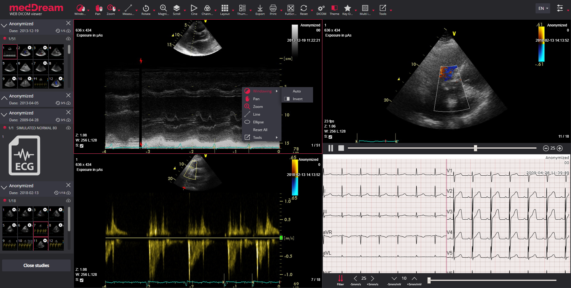
For ultrasound data MedDream also supports these features:

VTI. Velocity Time Integral used to measure the distance, which the blood was ejected over a date interval of time.

Up to 12 US studies may be opened at once.
DICOM ECG Viewer’s supported ECG devices:
Other parties’ brand names, with ownerships belonging to Mortara Instrument, custo med GmbH, SCHILLER AG, GE Healthcare, are not authorized by, sponsored by or associated with the MedDream trademark owner.
MedDream DICOM ECG Viewer can be to measure the volume of a 2D image by using Simpson’s approximation rule; the 2D area spun around a selected axis to form a 3D shape and the volume is measured. This technique allows volume measurements of a heart in a 2D computed radiography image.
MedDream DICOM ECG Viewer supports measurement of the Velocity Time Integral (VTI) on ultrasound (US) studies that can quantify the trace of the Doppler flow profile.
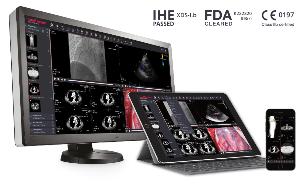
For more information, please read more about the MedDream WEB DICOM Viewer.
© 2007. Softneta. All rights reserved.